
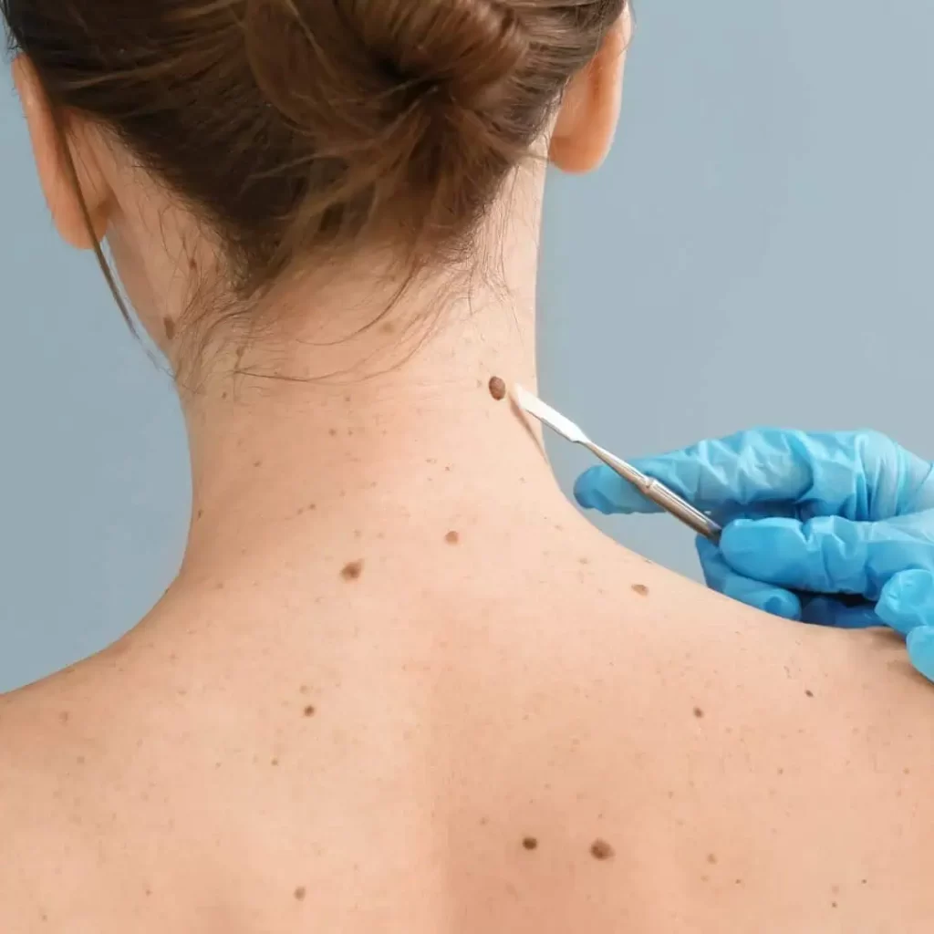
A free examination by a specialist in dermatology or cosmetic surgery should be scheduled if you notice any changes in the mole. That is, if it changes color, shape or size.
The same applies if the moles are in a place of constant irritation, are injured or itchy.
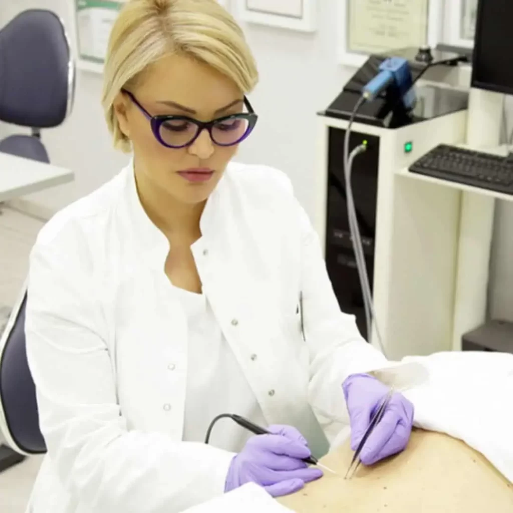
Based on the examination, our doctor assesses whether it is necessary:
- removal of moles by radio wave technique or
- by classical surgical method
with MANDATORY pathohistological analysis.
Of course, it is possible to schedule mole removal for aesthetic reasons as well.
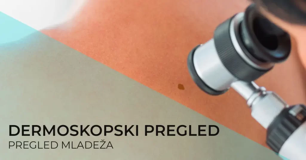
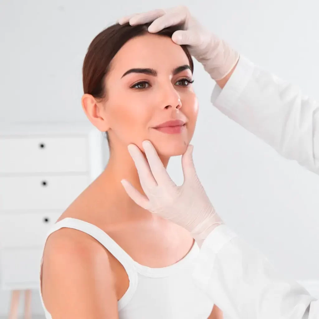
Free consultation for the removal of moles at the Diva Polyclinic
You can schedule a FREE consultation for mole removal in Belgrade with the doctors of the polyclinic Diva Dr. Bogdanović in one of our polyclinics or specialist dermatology offices:
- simple,
- painless,
- quickly feasible,
- non-invasive and
- very useful review of moles,
which helps specialist doctors to distinguish pigmentary changes on the skin from melanoma.
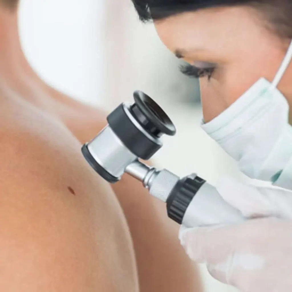
This diagnostic method is a preoperative examination that provides a three-dimensional image and provides insight into:
- structure of change,
- size,
- shape,
- shape and arrangement of blood vessels,
- the pattern and arrangement of pigment,
- presence or absence of regression structures or infiltration.
Diagnostics of pigment changes and skin diseases
- changes in blood vessels, as well as
- changes in keratinization and skin structure
with a diagnostic accuracy of 97 to 98%.
Timely and accurate diagnosis is crucial in the prevention of tumors.
The dermoscope creates a three-dimensional, high-resolution image.
Dermoscopy is necessary to monitor any pigmented lesion. With regular controls, especially in people with a large number of moles, the smallest change that occurs in the mole can be detected and removed in a timely manner.
Timely and accurate diagnosis is crucial in the prevention of tumors.
Why is regular dermoscopic examination of moles important?
It is estimated that the reliability of clinical examination of moles (medical examination with the “naked eye”) is about 65%. Dermoscopic examination of moles increases diagnostic accuracy up to 98% and therefore plays an important role in the early detection of melanoma and other skin tumors.
Reliable differentiation of benign from malignant changes
A clinical examination rarely provides insight into the shape and structure of blood vessels. This is often crucial for distinguishing benign from malignant changes, as well as for deciding whether it belongs to the group of benign changes, such as moles and fibromas, viral warts, or to the group of malignant changes, melanoma, basal cell or squamous cell carcinomas, for which timely and radical treatment is necessary.
What changes can be diagnosed using a dermoscopic examination?
In addition to reliably distinguishing benign from malignant changes, dermoscopy enables early detection of melanoma and mole changes.
Melanoma – skin cancer that has a high mortality rate, if diagnosed in late stages. Early diagnosis is crucial for patients, which was not possible before the use of dermoscopic devices. For example, in the countries of Western Europe today, around 80% of melanomas are detected at an early stage by dermoscopic examination, which significantly increases the chances of cure. After the suspicion of melanoma has been established, after a dermoscopy, the change on the skin is removed, and the final diagnosis is made after histopathological analysis.
Moles – self-examination of moles is recommended every 3 months, and dermoscopic examination of moles is recommended by a doctor, due to the increasingly frequent occurrence of skin tumors.
Special caution for risk groups
Dermoscopic diagnostics is extremely important for all patients who notice any changes on their moles, and especially for high-risk groups of patients such as:
- patients who have more than 50 moles,
- patients who have dysplastic or atypical moles,
- patients older than 50 years, who have severe skin damage caused by the influence of the sun,
- patients who often and a lot are exposed to the sun, who engage in risky professions, everyone who works outdoors – farmers, sailors and others,
- patients with a family history of skin cancer or melanoma,
because this method allows examination of the entire surface of the skin and follow-up for 3, 6 or 12 months, according to the need and dynamics determined by the specialist doctor.
People who belong to one of these high-risk groups should have dermoscopic examination of moles regularly.
Risk factors for skin tumors:
- family predisposition,
- excessive exposure to the Sun and UV rays in the solarium,
- people with light blonde or reddish brown hair,
- a large number of moles on the skin,
- light complexion and the presence of freckles.
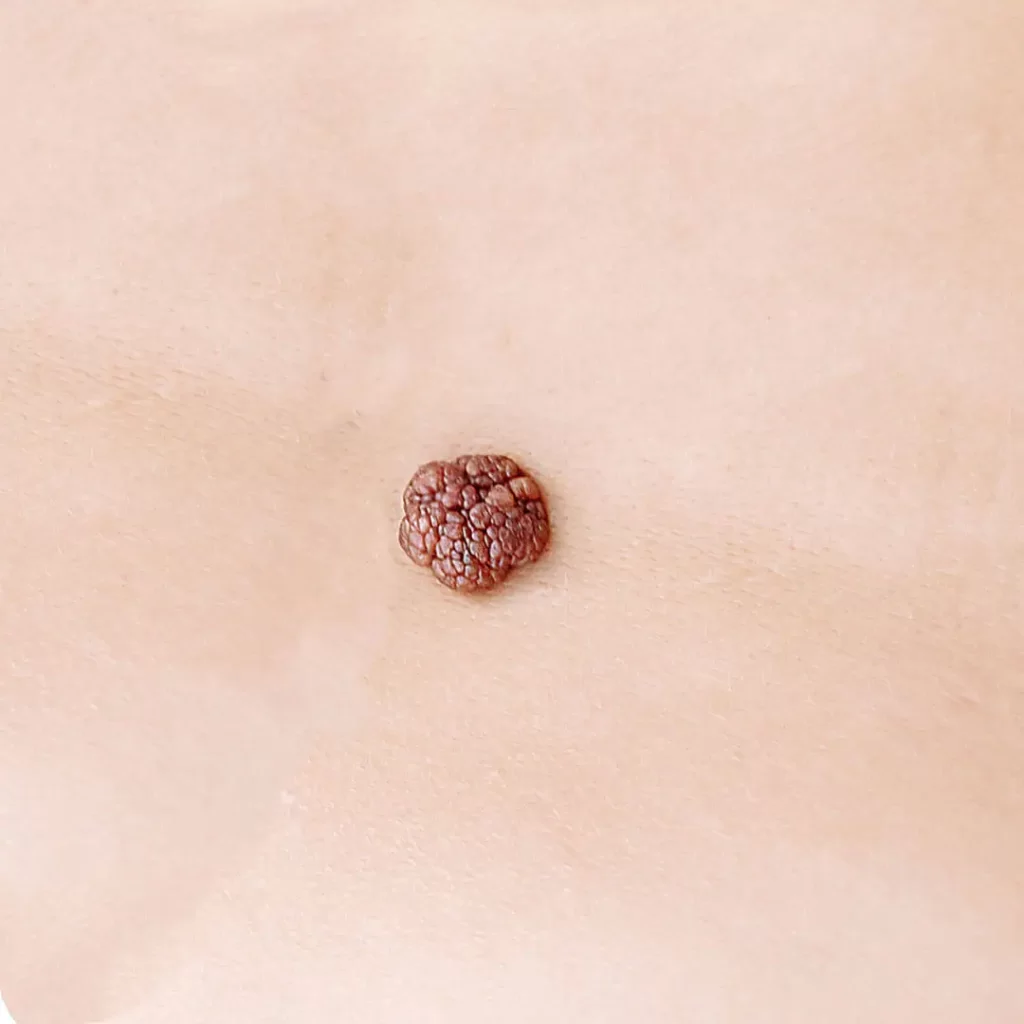
Which moles need to be examined with a dermoscopic device?
If any of the changes on the moles are observed, a dermoscopy is required. If the youth:
- grows rapidly,
- changes shape (asymmetry occurs),
- changes color,
- changes the edges (they become ragged and irregular),
- gets more different colors,
- rises above skin level over time…
Which other moles need to be monitored dermoscopically?
Youth who are atypical in appearance, even when melanoma is not suspected. They are also followed for 3, 6 or 12 months, after which dermoscopic images are compared and a decision is made as to whether they will be removed, followed by a histopathological analysis.
Dermoscopy is also recommended for monitoring congenital birthmarks, especially if they are larger than 10 centimeters in diameter.
Dermoscopy of moles before intervention - doctor's advice
Given that an increasing number of tumors are recorded, the doctor’s recommendation is to perform a dermoscopic examination of moles at least once a year. Warts, moles and other growths on the skin can represent potential foci of malignant tumors, so preoperative diagnosis with a dermoscopy device is extremely important, as it is crucial to rule out malignancy. In this way, the doctor will be able to make a decision in time whether the mole needs to be removed and which technique would be adequate to remove the mole.
What methods can be used to remove moles and other skin changes?
Depending on the type and size of skin changes and moles, they can be removed by classical surgical excision or by the radio-wave surgical method, with mandatory histopathological verification.
For prices for removing other skin changes, see our PRICE LIST.
NOTE:
Whether radio waves are an adequate method for removing moles will be assessed by a specialist doctor during a free consultation.
The results of the procedure may vary from patient to patient, and this is influenced by factors such as skin type, i.e. whether it is prone to scar tissue formation.
You can read the entire disclaimer HERE.
Appointment for a free mole examination or mole removal procedure in Belgrade
At the polyclinic Diva Dr. Bogdanović Belgrade, with our doctors specializing in dermatology and aesthetic surgery, you can schedule a FREE consultation for examination and removal of moles in one of our polyclinics or specialist dermatology offices:
to one of the phone numbers 063 338 334 or 011 3242 841 or to the email info@divaclinic.com
You can schedule an examination or procedure
· every working day from 9 a.m. to 8 p.m
· and on Saturdays from 9 a.m. to 3 p.m.
Price for specialist dermatological skin examination and dermoscopic examination of moles and other skin changes
If a specialist dermatological examination of the skin is required due to dermatological changes on the skin, then the price is RSD 6,000.
If you want to schedule only a dermoscopic mole examination, the price is 6,000 RSD.
Your “Diva Polyclinic”
Author of the text: Dr. spec. Svetlana Bogdanović
Co-author: Ph.D. Slavica Peković