
What are the safe methods to remove moles?
Our position and clinically proven medical fact is that the only reliable methods for removing moles are exclusively surgical techniques with MANDATORY pathohistological analysis:
- radio wave technique or
- classic surgical excision
When should a mole examination be scheduled?
After a clinical and, if necessary, a dermoscopic examination, the doctor decides whether the mole needs to be removed and which method is optimal for mole removal in the specific case. A big misconception is that moles should not be removed. It’s just the opposite!
They can be removed completely safely for practical reasons (when they are in places where they are exposed to friction or injury) or to improve the aesthetic appearance of the face or body. And preventive removal of moles is an absolute recommendation if they show changes that are risky for the patient’s health.
What is the optimal method for mole removal that is comfortable for the patient, with the maximum aesthetic result?
More than 90% of moles can be removed with radiosurgery. This includes all healthy moles, irritated or mildly altered.
The radio wave technique is a very advanced dermatosurgical method due to:
• speed and efficiency,
• high comfort for the patient,
• quick and easy healing
• very sophisticated, almost imperceptible scars.
Due to all these advantages, in most cases, it significantly suppresses the classic surgical removal of moles, as well as the laser removal of other changes on the skin, regardless of whether it is the removal of moles, warts, keratoses, fibromas, sebaceous cysts or xanthelasma from the skin of the face and bodies.
Radio wave mole removal experience
Due to all these advantages, in most cases, it significantly suppresses the classic surgical removal of moles, as well as the laser removal of other changes on the skin, regardless of whether it is the removal of moles, warts, keratoses, fibromas, sebaceous cysts or xanthelasma from the skin of the face and bodies.
Patient experience with radio wave mole removal
Our long-term patient Aleksandar S. shared his experience with radio wave removal of moles, who says:
“I have been to Diva Polyclinic twice so far and I am very satisfied.” Both times I came because of moles, which I have a lot of. One particularly bothered me and I managed to injure and hook it several times, so after the examination, in agreement with the doctor, I decided to remove it. It was painless and quickly over. The second time I came to the Diva polyclinic, I was very worried about a change in my skin that I had never had before. The doctor performed a detailed examination, talked with me, reassured me and advised me on how to protect my skin and moles from the sun. I sincerely recommend Diva Polyclinic for its expertise, knowledge and dedication to patients!”
Removal of healthy moles is recommended if they belong to the risk group. That is why radio-wave removal of moles is recommended as a preventive procedure.
Radiowave removal of moles - 12 ADVANTAGES
This distinguishes the radio wave method:
VERY PRECISE METHOD
Radio waves enable very precise removal of moles, which reduces the risk of damage to the surrounding tissue.
VERY PRECISE METHOD
Radio waves enable very precise removal of moles, which reduces the risk of damage to the surrounding tissue.
VERY PRECISE METHOD
Radio waves enable very precise removal of moles, which reduces the risk of damage to the surrounding tissue.
VERY PRECISE METHOD
Radio waves enable very precise removal of moles, which reduces the risk of damage to the surrounding tissue.
VERY PRECISE METHOD
Radio waves enable very precise removal of moles, which reduces the risk of damage to the surrounding tissue.
VERY PRECISE METHOD
Radio waves enable very precise removal of moles, which reduces the risk of damage to the surrounding tissue.
VERY PRECISE METHOD
Radio waves enable very precise removal of moles, which reduces the risk of damage to the surrounding tissue.
VERY PRECISE METHOD
Radio waves enable very precise removal of moles, which reduces the risk of damage to the surrounding tissue.
VERY PRECISE METHOD
Radio waves enable very precise removal of moles, which reduces the risk of damage to the surrounding tissue.
VERY PRECISE METHOD
Radio waves enable very precise removal of moles, which reduces the risk of damage to the surrounding tissue.
VERY PRECISE METHOD
Radio waves enable very precise removal of moles, which reduces the risk of damage to the surrounding tissue.
VERY PRECISE METHOD
Radio waves enable very precise removal of moles, which reduces the risk of damage to the surrounding tissue.
This distinguishes the radio wave method:
Why do the specialist doctors of the Diva polyclinic choose radiowave removal of moles as the optimal method?
Due to all these advantages, in most cases, it significantly suppresses the classic surgical removal of moles, as well as the laser removal of other changes on the skin, regardless of whether it is the removal of moles, warts, keratoses, fibromas, sebaceous cysts or xanthelasma from the skin of the face and bodies.
It is a modern advanced surgical technique and is one of the most popular because it gives GREAT results.
Moles, warts, keratoses, atheroma, xanthelasma, condyloma… are growths on the skin for which radio wave removal is very often the method of choice.
Dysplastic nevi, congenital nevi and all suspicious changes in the Diva polyclinic are performed exclusively by the classic surgical method, while other skin changes can be removed with radio waves.
The expert team of the Diva polyclinic, based on the examination, makes a decision whether the mole needs to be removed and whether the removal of the mole will be performed using the radio-wave technique or the classic surgical method with the use of local anesthesia.
When the mole is removed (regardless of which of the two mentioned methods it is removed), it must be sent for a histopathological examination in order to ensure the health safety of the patient.
The radio wave method of removing growths from the skin is widely used in plastic surgery and dermatosurgery. This is because in those branches of medicine it is very important to get as fine and almost imperceptible a scar as possible.
The advantages of radio wave surgery, when it comes to healthy moles, are numerous. This is why this is often the optimal method of choice when it comes to mole removal.
Why do the specialist doctors of the Diva polyclinic choose radiowave removal of moles as the optimal method?
You can schedule a FREE consultation for mole removal in Belgrade with the doctors of the polyclinic Diva Dr. Bogdanović in one of our polyclinics or specialist dermatology offices:
- DIVA Polyclinic Dr. Bogdanović - Kosovska 34 - Stari Grad Belgrade,
- DIVA Estetic Slavija, Vračar - Njegoševa 19a, Belgrade,
- DIVA Dermal New Belgrade - 125d Zorana Đinđić Boulevard, Belgrade.
Along with a free clinical examination, it is sometimes necessary to perform a mole dermoscopy in order to make a more precise preoperative diagnosis.
The price of demoscopy is 6,000 dinars.
Why is Diva polyclinic the right place for radio wave removal of moles and other skin changes?
What else, apart from removing moles, can radio wave removal be used effectively?
The radio wave technique is optimal for all benign skin changes, such as:
• Fibromas – very common growths on the skin made of collagen fibers and spindle cells, most often on the stalk.
• Viral warts – skin changes caused by different strains of HPV – human papilloma virus.
• Papillomas – hanging warts of viral origin.
• Keratoses – are caused by the formation of seborrheic keratin deposits.
• Sebaceous cysts – atheroma – epidermal cysts that are caused by blockage of the sebaceous glands’ outlet ducts.
• Xanthelasma – are formed on the eyelids in the form of yellowish deposits that can be flush with the skin or slightly raised.
• Condylomas – genital warts caused by human papilloma viruses.
• Dermatofibromas – benign growths flush with the skin, hard to the touch and most often occur on the hands and feet.
• Angiomas – better known as “red moles”, represent vascular changes and have no similarities in structure with moles.
• Granulomas – in the majority of cases, they are caused by inadequate and prolonged healing of wounds, resulting in the swelling of connective tissue with a tendency to rapid growth and a tendency to bleed.
• Syringomas – changes on the eyelids that look like small watery cysts, and arise as a result of blockage of the sweat glands.
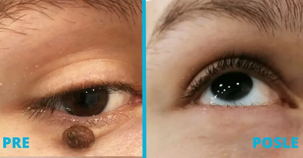
Radiowave removal of moles before and after the procedure
Does a scar remain after radio wave mole removal?

Radiowave removal of moles before and after the procedure
Young people are being removed from
• medical,
• aesthetic, but also
• practical reasons.
All three reasons are justified provided that the intervention is performed correctly.
Removal of moles, especially those that have undergone changes, is even recommended. But it is very important to determine an adequate method with mandatory PH analysis.
If the changes on the mole are discrete and dermoscopically do not show changes characteristic of skin tumors, they can be removed using the radio wave method with PH analysis.
However, if the clinical dermoscopic finding indicates an initial malignant alteration of the mole, it is MANDATORY to perform a wide surgical excision of the change with a detailed PH and immunohistochemical analysis.
Removal of moles and why pathohistological analysis is MANDATORY?
After it has been removed, at the Diva Polyclinic, the mole is ALWAYS sent for a pathohistological examination (PH), regardless of whether the intervention was performed with radio waves or a classic surgical technique.
Although the examination is performed by a specialist doctor before removing the mole, the cells can only be checked with certainty under a microscope and the presence of tumor cells can be ruled out.
In this way, by examining all the cells under a microscope, it is established what exactly the change is. And all with the goal of maximum patient safety.
Specialists in aesthetic surgery and dermatology at the “Diva” polyclinic perform a FREE consultation. That’s because we think it’s very important to react in time. And regular examinations of moles enable timely diagnosis of the resulting changes. Because when they are in the initial phase, then it is possible to cure malignant skin tumors with a simple surgical excision.
What is pathohistological analysis of moles and why is it important after a mole removal procedure?
In the pathohistological analysis process, the removed mole is observed under a microscope to determine the correct diagnosis.
Pathohistological analysis precisely determines what kind of skin change is involved. In addition, the composition of the change, its structure, growth rate…
There are two parts to the pathohistological examination:
• microscopic (includes histological and cytological analysis) i
• molecular-biological (determines the characteristic features of the resulting changes).
Of course, before the removal of the mole at the Diva polyclinic, it is examined by a specialist doctor. However, only under a microscope can the cells be checked with certainty and the presence of tumor cells can be ruled out.
The most common reasons for mole removal?
The most common reasons patients come in for mole removal is when:
• for aesthetic reasons, when they are on the face, neck or décolleté
• for practical reasons when located in places where they are exposed to irritation or injury or
• if they are on the hairy part of the head – hair, in the hair, and can be injured during combing,
• the mole shows signs of rapid growth.
Or when the change has already occurred, i.e. when the youth:
• changes color,
• shape,
• itching,
• stings,
• bleeding
• or is in a place where it is often injured,
then an examination by a specialist and preventive removal is recommended.
What does mole removal look like at the "Diva" polyclinic?
A clinical diagnosis is made during the clinical examination at the “Diva” polyclinic.
And if necessary, a dermoscopic examination is also performed. In this way, an in-depth examination of the mole is performed, and the method is
• simple,
• painless and
• non-invasive.
Dermoscopic examination of moles before the mole removal procedure
When should a mole examination be done? Is it only done before the mole removal procedure?
Radiofrequency mole removal success in numbers
After more than 25 years of clinical experience and follow-up of patients to whom we performed mole removal with radio waves, this is our conclusion:

the mole can be removed with radio wave surgery

patients are satisfied with the aesthetic result

mole removal in one intervention
When is mole removal necessary?
Based on the examination, the expert team of the “Diva” polyclinic decides whether an intervention is necessary or not. In addition, a decision is made whether the removal of the mole will be performed using the radio-wave technique or the classical surgical method with the use of local anesthesia.
Who performs mole removal at the "Diva" polyclinic?
Both mentioned mole removal procedures at the “Diva” polyclinic are performed exclusively by doctors specializing in plastic surgery and dermatology with more than 25 years of experience.
What are all the methods for removing moles?
There are different ways to remove moles, however NOT ALL METHODS ARE SAFE for the patient.
Techniques for removing moles RECOMMENDED by the expert team of the Diva polyclinic
Given that the patient's health is our first priority, we only do these two methods at the Diva Polyclinic:
• radiowave dermatosurgical technique,
• classic surgical method - mole excision.
After these medical procedures, IT IS POSSIBLE TO SEND THE YOUTH FOR PATHOHISTOLOGICAL ANALYSIS!
Mole removal techniques that we ABSOLUTELY DO NOT RECOMMEND!
Other techniques used to remove moles that WE ABSOLUTELY DO NOT RECOMMEND because they are unsafe for patients are:
• removal of moles with an erbium or CO2 laser,
• mole removal with electrocautery.
• shave biopsy,
• treatment with different acids.
When using these methods, it is NOT POSSIBLE to perform a pathohistological examination of mole cells.
When is it time to remove moles - the ABCDE rule?
After more than 25 years of clinical experience and follow-up of patients to whom we performed mole removal with radio waves, this is our conclusion:
ASYMMETRY
When ASYMMETRY is present. Benign moles are mostly symmetrical, regardless of their shape - oval or round. If the mole is irregularly shaped and asymmetrical, of unequal height, larger or thinner on one part, the advice is to contact a specialist doctor who will perform an examination.
BORDER
If the edge of the mole is not regular, but jagged - BORDER (eng), it is recommended to consult a doctor.
COLOR
If the colors inside the mole are different or it suddenly changed color - COLOR (eng). Juveniles are usually a uniform shade of brown. When the mole has several shades of brown, black and blue, it is necessary to perform an examination.
DIAMETER
If the mole suddenly expands, i.e. increases in diameter - DIAMETER (eng), make an appointment with a specialist.
EVOLVING
If a mole suddenly starts to grow or suddenly changes in some other way - EVOLVING (eng), see a dermatologist.
None of the above changes, if noticed in time, are alarming.
Appointment for a free mole examination or mole removal procedure in Belgrade
At the polyclinic Diva Dr. Bogdanović Belgrade, with our doctors specializing in dermatology and aesthetic surgery, you can schedule a FREE consultation for examination and removal of moles in one of our polyclinics or specialist dermatology offices:
to one of the phone numbers 063 338 334 or 011 3242 841 or to the email info@divaclinic.com
You can schedule an examination or procedure
· every working day from 9 a.m. to 8 p.m
· and on Saturdays from 9 a.m. to 3 p.m.
Price for specialist dermatological skin examination and dermoscopic examination of moles
If a specialist dermatological examination of the skin is required due to dermatological changes on the skin, then the price is RSD 6,000.
If you want to schedule only a dermoscopic mole examination, the price is RSD 6,000.
Your “Diva Polyclinic”